Map the meaning of your gum spots and patches
You are brushing your teeth, leaning a little closer to the mirror, when something catches your eye. A small patch on your gums appears different from the surrounding tissue. It might look white and slightly textured, bright red and irritated, or dark like a tiny freckle.
Moments like this often trigger immediate concern. Any unfamiliar change inside the mouth can feel unsettling.
In reality, the gums can display a wide variety of colours and patterns. Some marks are harmless variations in pigmentation. Others reflect irritation, inflammation, or an underlying oral condition. In rarer cases, a patch may signal something that requires prompt professional attention.
Understanding what these changes might represent can remove unnecessary worry while helping you recognise when a dental examination is important. The colour, texture, and duration of a patch often provide useful clues about what may be happening beneath the surface.
Why do gums suddenly look different
The gums are living tissue that constantly respond to their environment. Everyday factors that change within your mouth, such as temperature, bacteria, minor trauma, and even hormonal changes, influence their appearance.
Patches or spots may develop due to:
- Local irritation from sharp foods, dental appliances, or aggressive brushing
- Bacterial or fungal infections
- Inflammatory conditions affecting the oral tissues
- Natural pigmentation caused by melanin
- Medication reactions or systemic conditions
- Tobacco exposure
Most of these causes are manageable and often temporary. The key lies in observing how the patch behaves over time and whether additional symptoms appear.

White patches
White areas on the gums can appear as small plaques, thin films, or delicate lace-like patterns. These changes are often associated with alterations in the surface layer of the oral tissue.
One common cause is leukoplakia, a condition where the tissue thickens in response to chronic irritation. Tobacco use, friction from dental appliances, or persistent inflammation may contribute to its development. Leukoplakia patches are typically firm and cannot be wiped away.
Another possibility is oral lichen planus, an inflammatory condition that may create intricate white lines across the gums or inner cheeks. While usually benign, it can cause sensitivity or discomfort when eating spicy or acidic foods.
Fungal infections such as oral thrush may also produce white patches. Unlike leukoplakia, thrush often forms creamy deposits that may rub away, leaving slightly red tissue underneath.
White patches should always be assessed by a dental professional if they persist. Many are harmless, yet a thorough examination ensures that more serious conditions are not overlooked.
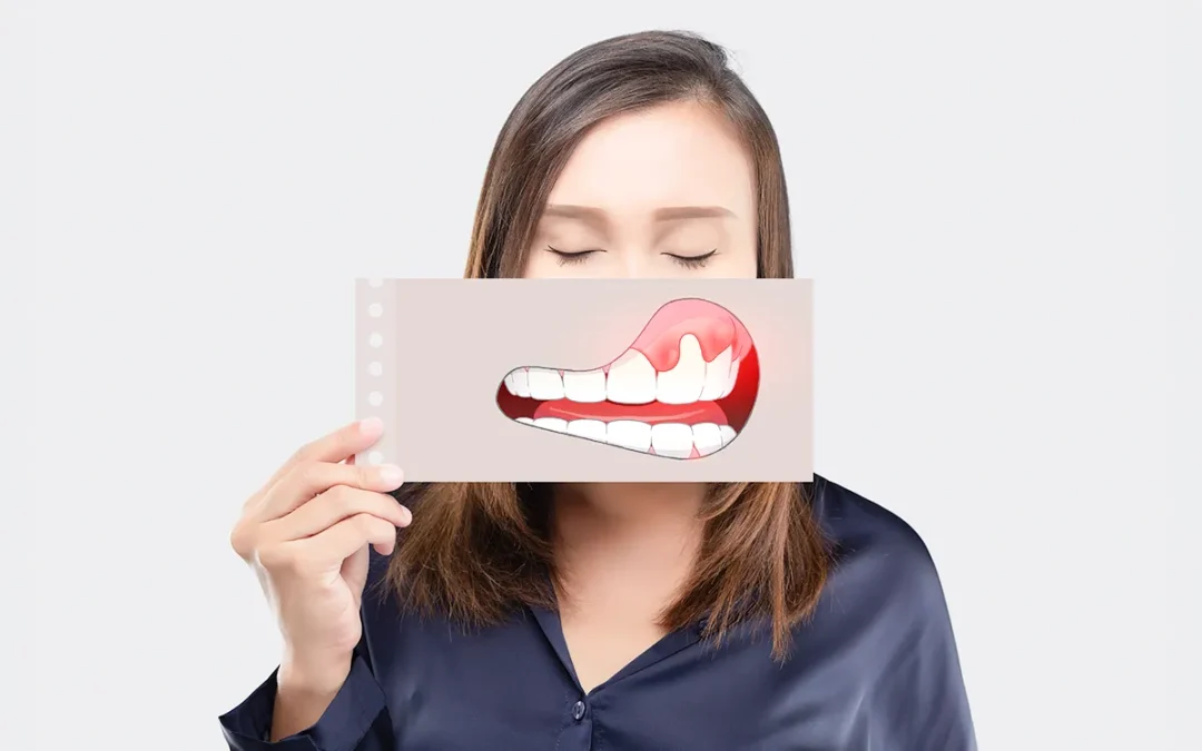
Red patches
Red patches often indicate inflammation or thinning of the gum tissue. These areas may appear smooth, slightly shiny, or unusually bright compared with surrounding gums.
Irritation from dental plaque is a frequent cause. When bacteria accumulate along the gumline, the tissue becomes inflamed, producing redness and tenderness. Improved oral hygiene and professional cleaning often resolve the issue.
Some red patches, however, require closer attention. A condition known as erythroplakia produces vivid red areas that cannot be explained by simple irritation. Although uncommon, these lesions carry a higher potential for cellular changes and should always be evaluated promptly.
Redness may also occur with traumatic ulcers, burns from hot food, or allergic reactions to oral products.
Persistent red patches that do not heal within a short period deserve professional assessment.

Brown, black, or blue marks
Dark spots on the gums often resemble freckles. In many individuals, particularly those with naturally higher melanin levels, these areas represent normal pigmentation.
These pigmented spots may appear:
- Flat and evenly coloured
- Symmetrical across the gum tissue
- Stable in size over time
Dental materials can also produce dark marks. For example, small bluish-grey areas sometimes develop near older metal fillings. These are known as amalgam tattoos and are generally harmless.
Occasionally, darker lesions require careful evaluation. Rare conditions such as oral melanoma can begin as a pigmented spot that gradually changes in colour, shape, or size. Such cases are uncommon but reinforce the importance of monitoring new or evolving patches.
Any pigmented area that grows, changes appearance, or develops irregular borders should be examined by a dentist without delay.
Ulcers, blisters, and temporary patches
Not all spots represent long-term conditions. The mouth frequently develops temporary lesions that heal naturally.
Mouth ulcers, also known as aphthous ulcers, are small painful sores that appear on the soft tissues of the mouth, including the gums. Stress, minor trauma, or immune responses may trigger them. Most resolve within one to two weeks.
Blister-like patches may also occur from burns, friction, or viral infections such as cold sores.
While these conditions often heal without intervention, frequent or unusually large ulcers should be discussed with a dental professional.

The 2-week rule
A simple guideline helps distinguish temporary irritation from something that requires professional evaluation.
Any spot, patch, or ulcer in the mouth that does not heal within two weeks should be examined by a dentist.
This timeframe allows the mouth’s natural healing processes to resolve minor injuries. When a lesion remains unchanged beyond this period, further investigation may be appropriate.
Dentists are trained to identify subtle variations in oral tissues. Early assessment ensures that potential concerns are addressed quickly and effectively.
What to expect during a dental examination
A dental assessment for gum patches is typically straightforward and comfortable.
The dentist will examine the area closely, looking at its colour, texture, borders, and location. Questions may include when the patch first appeared, whether it has changed, and whether any symptoms such as pain or bleeding are present.
Additional steps may involve:
- Reviewing medical history and medications
- Assessing oral hygiene and gum health
- Taking photographs to monitor changes
- Referring for further evaluation or biopsy if needed
Most cases require only observation and routine monitoring. When treatment is necessary, early detection often allows for simple and effective management.

Gum care in daily life
Healthy gums provide the foundation for a healthy smile. Daily care plays a significant role in preventing many conditions that cause patches or irritation.
Consistent habits support gum health:
- Brush thoroughly twice daily with a soft-bristle toothbrush
- Clean between teeth using floss or interdental brushes
- Attend regular dental examinations and professional cleanings
- Avoid tobacco products
- Maintain a balanced diet that supports oral health
These small routines help protect the delicate tissues of the mouth while reducing the risk of inflammation and infection.
Partnering with your dentist for long-term gum health
Not every spot on your gums is a problem, but every change deserves a second look. Most marks are harmless. Natural colour variations, mild irritation, temporary ulcers and they settle on their own. The difference is in what changes, what lingers, what doesn’t quite belong.
That’s where your regular dental check-ups come in. They enable your dentist to spot, monitor, and protect, guiding you to the right treatment or advice before problems escalate. Regular professional guidance, combined with diligent daily care, helps maintain strong gums and a confident smile. So before something catches your eye in the mirror, let your dentist take a closer look before it becomes something you can’t ignore.
